Our Vision Care Technology
Ziess Clarus 500
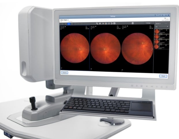
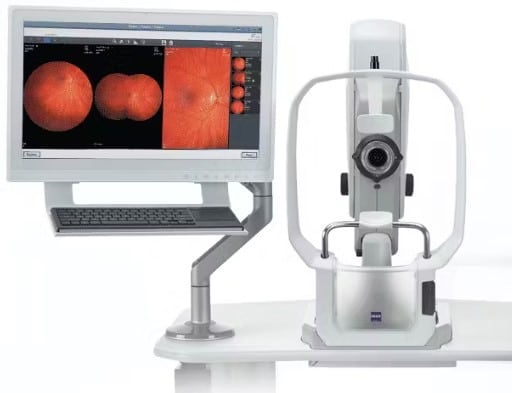
The advent of widefield retinal imaging has shown us that indications of disease are often located in the far periphery of the retina. CLARUS™ 500 is the next generation fundus imaging system from ZEISS that provides True Color and high-resolution across an entire ultra-widefield image.
True Color
ZEISS CLARUS 500, the newest ultra-widefield retinal camera from ZEISS, allows clinicians to use color to their diagnostic advantage. ZEISS CLARUS generates images that closely resemble the coloration of the fundus as seen during clinical examination. Color fundus imaging can aid in the diagnosis and documentation of ocular disease, ensuring confidence when evaluating the optic disc, nevi and lesions where color is important.
Exceptional Clarity
ZEISS CLARUS 500 captures clear and accurate images from the macula to the far periphery. Early indications of disease can often be subtle and difficult to see through direct observation or low resolution fundus imaging. Leveraging ZEISS optics, ZEISS CLARUS 500 is an ultra-widefield retinal camera that captures a high-resolution image down to 7 microns.
Comfort
Simple, stable and intuitive, ZEISS CLARUS 500 is an ultra-widefield retinal camera that has been purposefully designed to optimize each patient’s experience
The Live IR Preview in ZEISS CLARUS 500 allows the technician to optimize alignment – intervene with lid and lash, and remove image artifacts before capturing an image. The result is fewer image captures for the patient and a more efficient imaging process for the practice.
Fewer Recaptures
The Live IR Preview in ZEISS CLARUS 500 allows the technician to optimize alignment – intervene with lid and lash, and remove image artifacts before capturing an image. The result is fewer image captures for the patient and a more efficient imaging process for the practice.
Topcon Maestro 2 Robotic OCT and Color Fundus Camera
User-Friendly: Robotic OCT and fundus camera with single-touch automated capture
Widefield Scan: 12x9mm 3D wide scan captures macula and optic disc and includes the Hood Report for Glaucoma
High Resolution:
Multimodal Imaging: OCT and true color fundus photography.
Olleyes Virtual Reality Visual Field Testing
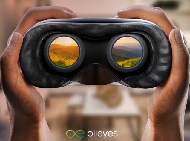
Patients using the VR headset can sit in any position they find comfortable — including lying down. Patients can move their head during testing. Room lighting can remain on. The device tests both eyes simultaneously. Extra time isn’t needed to switch between eyes during testing. This multi-test system includes acuity and color vision testing.
The headset weighs less than 7 ounces, fits over the patient’s head like any VR headset, and can be worn comfortably over glasses, so additional corrections aren’t needed.
HD Exam using our Digital Refraction System
Provides you as a patient with a comfortable experience. High Technology Digital Refraction provides vision testing with an HD monitor with a virtually unlimited combination of alphabet, number, character and pictures.
The Wavefront Auto Refractor/Keratometer / Aberrometer is the newest generation digital refraction system. This model combines the features of an auto refractor, auto keratometer and an aberrometer into one convenient-to-use instrument.
It is based on the Hartmann-Shack principle and focuses on measuring second-order aberrations, which result in myopia or hyperopia and regular astigmatism.
Because correcting second-order aberrations has the highest impact on acuity, which is the eye’s ability to distinguish object details and shape.
Electronic Records Interface – allows us to quickly interface the pre-test room results to the exam room to modify the prescription and to interface into the patient’s chart. Increases speed and accuracy of the exam process.
Wavefront Maps – leads better understanding for patients’ eye conditions since it graphically shows the second-order aberrations including myopia, hyperopia and astigmatism.
RMS (root mean squared) – Provides RMS to quantify the amount of aberration and describe the magnitude of an eye’s second order aberration for 3.0 mm pupil size. 2 mm
Minimum Pupil Size -. It only requires a 2mm pupil diameter to acquire accurate reading minimizing errors cause by eyelid interference.
Residual Astigmatism – Measures the refractive error of the total eye, corneal astigmatism and calculates Residual astigmatism. This valuable information provides more information in regards to the eye as an entire system. In addition the residual astigmatism values can be printed out.
Peripheral Keratometry – Capable of providing five directional keratometry readings, one central and four peripheral readings. This additional information offers enhanced data for better contact lens fitting.
Continuous Measurement Mode – While pressing and holding down the joystick button, continuous measurement can be completed without a delay to fog the patient’s vision. This provides quick and rapid measurements.
Visual Fields Automated Perimeter
Modern diagnostic instrument for precise and fast testing of field of vision.
Accurate / Intelligent
Depending on chosen test strategy, it enables defining the sensitivity threshold of retina in a given area, as well as making a fast screening test. Accurate, intelligent advanced threshold, Screening and quick 3 – zone can be selected according to needs and patient’s condition.
Saves Time
Advanced strategy which utilizes special algorithm allows quicker examination without loss in result resolution. 30-2 field can be tested in less than 3 minutes. Additionally 3 accuracy levels can be selected. “Reduced Field” and “Neurological Field Reduction” options help reduce examination time for other strategies.
Reliable
Examination reliability can be estimated on the basis of false negative and false positive tests. Built-in digital camera allows eye-detection during examination and during setting patient’s position. Thanks to pupil tracking system it allows a continuous automatic control of fixation. When patient loses fixation, blind spot is examined and special attraction mechanism turned on. Well known Heijl-Krakau method allows tracking of a blind spot and monitoring of examination reliability. All reliability indices are presented in examination results and on the printout.
Kinetic Test
Thanks to kinetic strategy it is possible to examine hardly cooperating patients with big defects for whom static examination is difficult to perform. 4 stimuli colors, I-V Goldmann sizes, adjustable stimuli velocity, silent operation, built-in programs together with option of retesting arbitrary vectors defined by user make PTS1000 a top product for kinetic perimetry.
Electronic Medical Records
EMR (Electronic Medical Records) is the digital documentation of a patients’ medical history and care. Your health and medical records are being kept, accessed, changed and updated digitally, using computers.
All current medical records at Music City Optical are electronic. There are many benefits for you.
If you are a previous patient, Dr. James will already have information about any medications you take, the results of any lab tests you’ve had, any health issues you’re facing, and prior prescriptions all in one convenient place.
The Doctor and staff at Music City Optical won’t have to pull charts and transcribe information, so they could possibly have more time and more meaningful interactions with patients.
Electronic records improve communication among members of the care team, as well as communication between you and Doctor James.
Electronic records allows Dr. James to concentrate even more on patients and not the paperwork.
EMR has organized and simplified the prescription process. Spectacle and contact lens prescriptions are available to anyone in the office at any time, increasing efficiency and accuracy. Eyeglass prescriptions are generated in the exam room and instantly become available to the frame stylist or optician, saving time and eliminating the possibility of transcription error.
Retinal photographs are integrated into the chart so that conditions can be monitored each year relying on the actual images of the eye instead of drawings.

